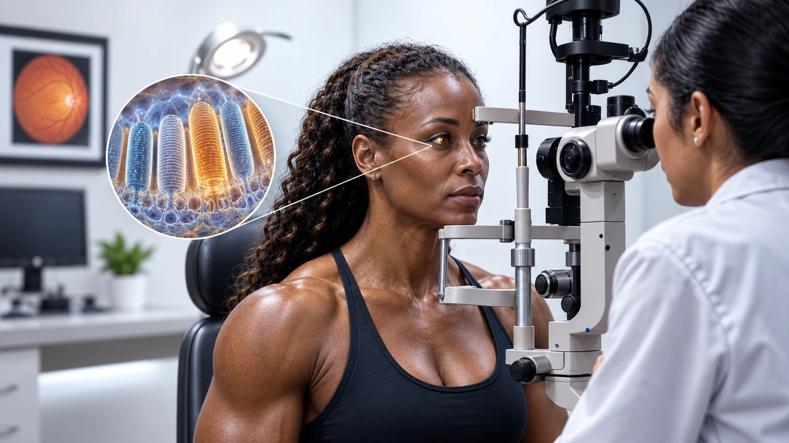

Omega-3 fats are often introduced as “brain nutrients,” but one of the most concentrated places to find docosahexaenoic acid—DHA—is the retina. In the outer segments of light-sensing photoreceptor cells, DHA can account for more than half of the phospholipid fatty-acid chains, depending on the membrane and measurement. That is a remarkable structural fact. It is not the same as proof that taking extra fish oil will sharpen a healthy adult’s vision.
The WorkoutHealthy Facebook post focuses on the first part of that story. This article explains the photoreceptor membrane, how it participates in turning light into a neural signal, what animal deficiency studies tell us, and where supplement claims outrun human evidence.
Where DHA is concentrated
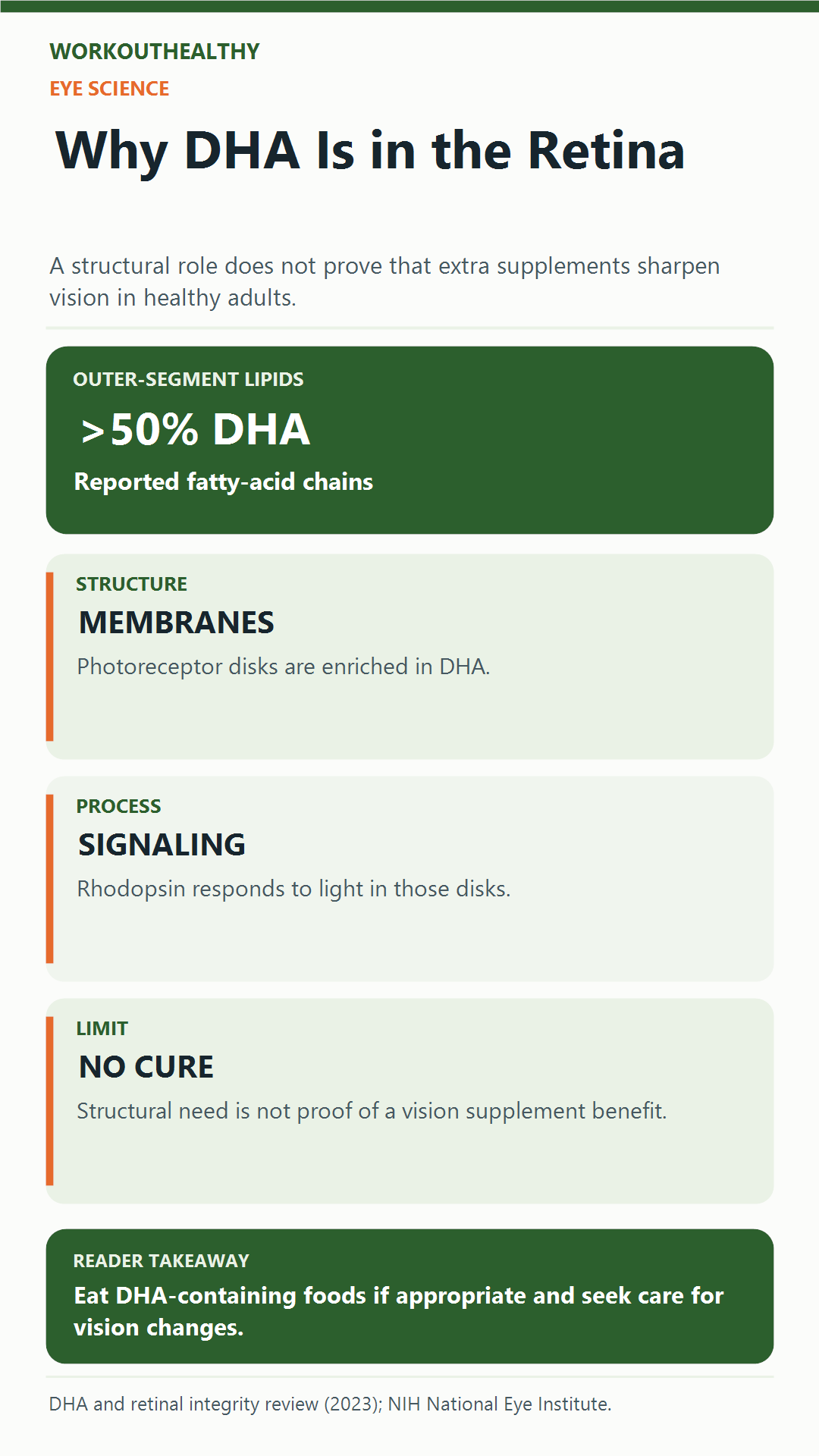
DHA is a long-chain omega-3 fatty acid with 22 carbon atoms and six double bonds. It is found in many neural tissues, but rod and cone photoreceptors are especially interesting because their outer segments contain stacks of membrane disks used to detect light. A 2023 review of retinal biology reports that DHA accounts for more than 50% of the fatty-acid side chains in photoreceptor outer-segment phospholipids. Exact percentages vary with tissue, species and analytical method, so “60% of every fatty acid in your entire retina” would be too broad.
Photoreceptors continually renew these disks. New disks form near the cell body, and older material is shed and cleared by the adjacent retinal pigment epithelium. DHA is one of the lipids incorporated into this highly specialized system. Reviews describe active uptake, sorting and conservation of DHA within retinal tissue. The body’s apparent preference for retaining DHA is part of why scientists consider it important—not a reason to assume that adding ever-larger doses brings ever-better sight.
The attached social creative shows an eye examination. It also includes glowing illustrations of other organs that should be read as design, not as a medical scan of the person depicted. Our article hero uses a more focused conceptual photoreceptor inset to reflect the actual subject. Neither image shows what the patient’s own retina contains.
A high concentration of a nutrient in a tissue tells you that the tissue uses it; it does not determine the right supplement dose.
How photoreceptors respond to light
Light reaches the outer segment of a rod or cone. There, visual pigment molecules—including rhodopsin in rods—begin a signaling cascade when they absorb light. That process, called phototransduction, changes the photoreceptor’s electrical activity and ultimately helps the visual system relay information toward the brain. The National Eye Institute places this conversion in the photoreceptor outer segment.
DHA-rich membrane environments can affect physical properties of those disks and the behavior of proteins embedded in them. Laboratory research has examined how membrane lipid composition changes rhodopsin packing and function. It is tempting to compress this into “DHA makes rhodopsin move faster,” but the actual biophysics is more complex. Membrane composition influences a network of protein states; the effects cannot be reduced to a single speed claim for daily vision.
This is the most important distinction in the Facebook post. DHA has a direct structural association with the machinery of vision. That is much more specific than a vague wellness slogan. Yet the retina’s multiple layers, nutrient transport, genetics, age and disease all influence the final visual outcome. A molecular role is not a clinical guarantee.
What deficiency studies show
Much of the strongest experimental evidence for DHA’s role comes from animal models. When animals are raised on diets deficient in omega-3 fats, retinal DHA levels can fall and electroretinogram responses can change. A review of retinal DHA biology describes that association while also noting that the precise mechanisms behind altered function are not fully resolved. Animal data are valuable for establishing a plausible need; they do not imply that every adult eating a typical diet is DHA-deficient.
Developmental stage matters. A growing visual system may have different needs from a mature one. It would be misleading to take a result from developing rats or from an unusual deficiency model and promise that an omega-3 capsule will correct a healthy adult’s night vision. The correct conclusion is narrower: DHA is a prominent component of photoreceptor membranes, and deficient conditions can disrupt retinal biology.
Another caveat is that dietary fats travel through digestion, circulation and tissue-specific transport before reaching the retina. The amount swallowed is not the amount that appears in a particular photoreceptor disk. This makes simplistic “one capsule equals one eye benefit” arithmetic impossible. Nutrition research has to measure real clinical outcomes—not just biological plausibility.
Deficiency evidence and supplementation evidence answer different questions.
Some posts describe the retina as “DHA-dense” without identifying the precise compartment. That wording is not wrong, but it can hide the scientific point. The extraordinary fraction is reported for particular photoreceptor outer-segment phospholipids, not for every cell, fluid or fatty acid in the whole eye. That specificity is why an anatomical explanation belongs in the article. It lets readers appreciate the finding without turning a subcellular measurement into a whole-body nutrition target.
It also helps to ask what a study actually measured. Chemical analysis can show the fatty-acid composition of a tissue. An electroretinogram can show an electrical response under experimental conditions. A visual-acuity test asks whether a person can resolve detail, and a disease trial asks whether outcomes change over time. Those measurements are related, but they are not interchangeable. A strong biological rationale for DHA motivates clinical research; it does not substitute for it. Keep those levels separate when evaluating any claim about an eye supplement.
What supplements have not proven
DHA-containing foods can be part of a healthy diet, and supplements have been studied for several eye conditions. But “the retina contains DHA” is not itself a trial result showing that extra DHA improves sight in a well-nourished person. For dry eye disease, a Cochrane review of omega-3 and omega-6 trials described evidence as uncertain and inconsistent overall. Dry eye is also different from retinal photoreceptor function, so its results should not be repackaged as a retinal cure.
Specific eye diseases require specific evidence. Macular degeneration, diabetic retinopathy, inherited retinal conditions, dry eye and ordinary refractive errors have different mechanisms and treatments. A supplement marketed broadly for “eye health” may not address the condition a person has. If your vision changes, particularly suddenly, a qualified eye-care professional—not a nutrition headline—should guide evaluation.
High-dose fish oil may also be inappropriate for some people, depending on medicines, bleeding risk, cardiovascular history and the product’s dose. More is not automatically safer or better. Ask a clinician before using concentrated supplements for a medical goal, especially if you already have an eye diagnosis. Our guide to fitness supplements and evidence explains why a clear outcome matters more than a persuasive mechanism.
Food and practical eye care
Common dietary sources of DHA include fatty fish, and algae-derived products offer an option for people who avoid fish. Fish also provides other nutrients, while species and serving frequency matter for mercury exposure. Our SMASH fish guide covers the benefits and limitations of simple fish-choice rules. A balanced dietary pattern is a more defensible starting point than trying to “max out” one nutrient because it appears in a particular organ.
For everyday vision care, regular eye examinations, addressing changes promptly, managing known conditions such as diabetes, and following appropriate clinician advice are not replaced by omega-3 intake. Sunglasses, smoking avoidance and blood-pressure management may matter depending on the condition. The exact plan should come from your eye-care professional, particularly if you have symptoms.
Think of DHA as one structural ingredient in a complex biological system. A building needs materials, but pouring more material on the roof does not automatically improve its function. The analogy is imperfect, but it captures the main caution: adequacy and clinical benefit are not interchangeable. If you already eat a varied diet and have no demonstrated deficiency, a supplement claim needs direct evidence that it improves the outcome you care about.
Bottom line
DHA is genuinely abundant in photoreceptor outer-segment membranes, where light detection begins. Reviews report that it can make up more than half of certain membrane fatty-acid chains, and animal deficiency studies link low retinal DHA with altered visual responses. Those findings establish a meaningful structural role. They do not prove that extra DHA sharpens vision or treats an eye disease in a healthy adult. Choose food sources that fit your diet, evaluate supplements against disease-specific clinical evidence, and seek professional care for vision changes. This article is educational, not medical advice.
References
- The essential role of docosahexaenoic acid and its derivatives for retinal integrity. 2023 review. PubMed.
- The role of docosahexaenoic acid in retinal function. 2001 review. PubMed.
- National Institutes of Health, National Eye Institute. Photoreceptor outer segments and phototransduction. NIH.
- Effect of dietary DHA on rhodopsin content and packing in photoreceptor membranes. PubMed.
- Cochrane review: omega-3 and omega-6 supplements for dry eye disease. Cochrane.