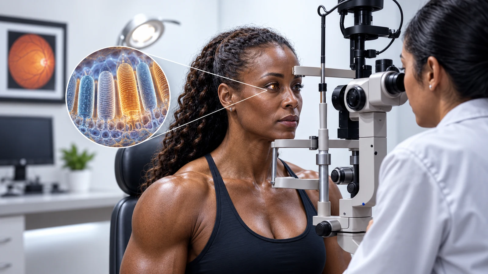

Illustration note: The eye-examination scene and photoreceptor inset are conceptual artwork, not an identified patient, a retinal scan or a measurement of anyone’s DHA level.
DHA is highly concentrated in particular retinal membranes. That helps explain its biological importance, but it does not tell us that taking extra DHA will sharpen a healthy adult’s vision.
The useful question is where the DHA is, what experiments show it doing and whether a human trial tested the benefit being advertised. Those are three separate steps in evaluating a DHA retina claim.
This guide follows the evidence from photoreceptor membranes to clinical outcomes. It also explains why a trial’s primary electrical-response result and a later visual-field analysis can produce different headlines without becoming two independent trials.
Where the “more than 50%” retinal figure belongs
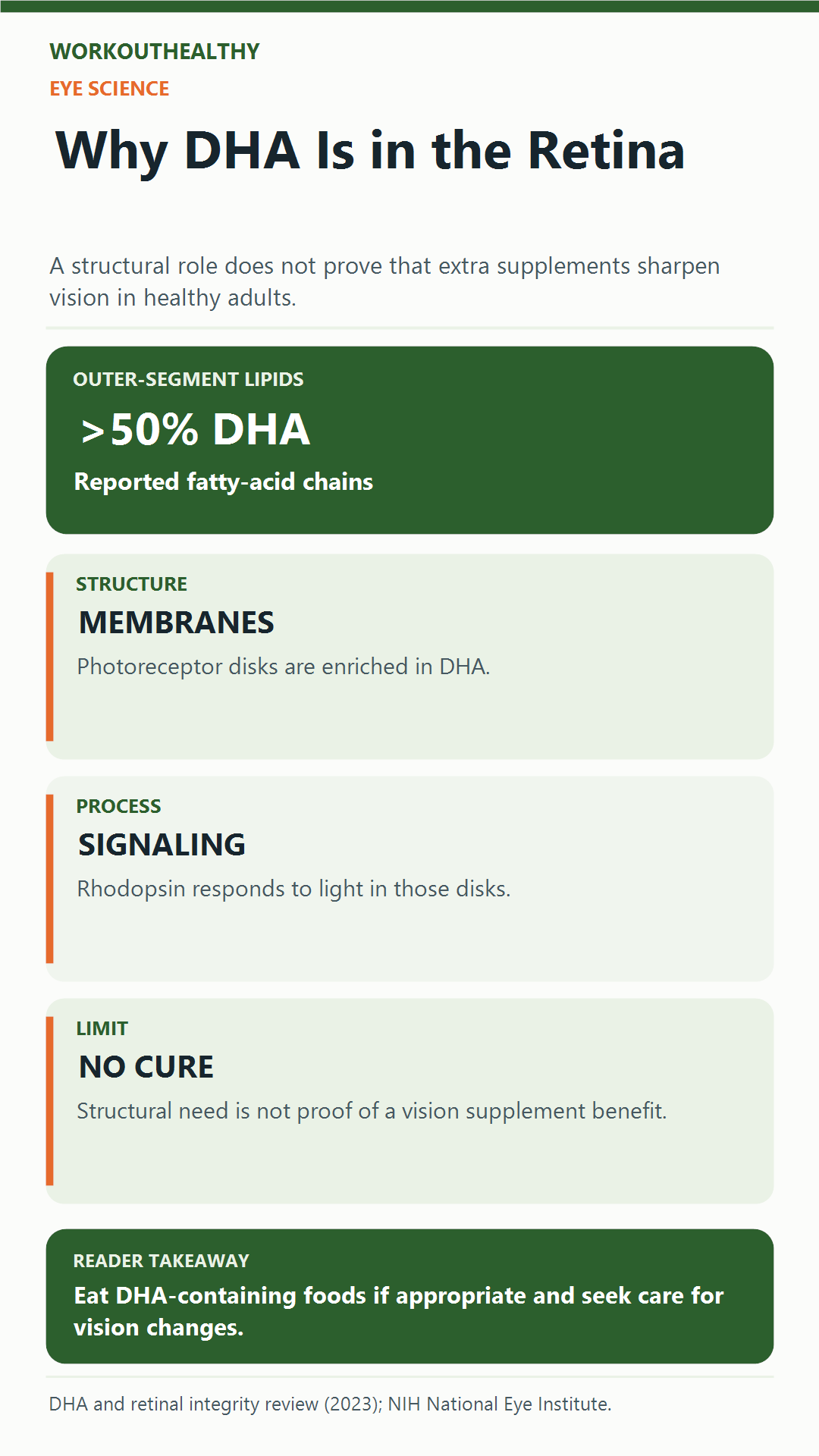
A 2023 review by Swinkels and Baes describes DHA as accounting for more than 50% of the fatty-acid side chains in photoreceptor outer-segment phospholipids. The denominator is a particular membrane component, not the weight of the whole retina.
Reading the anatomy from the outside in makes that distinction easier:
- Retina: the light-sensitive tissue at the back of the eye, containing multiple cell types.
- Photoreceptor: a rod or cone, specialized for detecting light.
- Outer segment: the photoreceptor region containing the membranes involved in light detection.
- Phospholipid: a membrane-building molecule with fatty-acid chains.
- DHA: one of those fatty acids, especially enriched in this membrane compartment.
“More than half of these fatty-acid chains” therefore does not mean that half your eye is DHA. It also cannot be converted into a supplement percentage or a daily milligram target.
The exact composition varies with the tissue compartment and measurement. A careful description keeps “photoreceptor outer-segment phospholipids” attached to the striking number.
What membrane experiments actually demonstrate
DHA-containing phospholipids help provide the physical environment in which photoreceptors work. Rhodopsin, the light-sensitive pigment in rods, operates within that membrane system.
A 2017 experimental study by Shindou and colleagues investigated an enzyme called LPAAT3, involved in incorporating DHA into phospholipids. In mice lacking that enzyme, DHA-containing phospholipids were greatly depleted in photoreceptor outer segments.
The mice had disordered membrane-disc structure and impaired electroretinogram responses. Laboratory membrane experiments and simulations also supported a role for DHA-containing lipids in membrane flexibility.
That is meaningful evidence about retinal structure and function in the model studied. It is not a randomized trial showing that a capsule improves ordinary eyesight in people with adequate nutrition.
The distinction matters because an enzyme-deletion model is more specific than simply feeding an otherwise healthy person a little less DHA. Neither an experimental mutation nor an animal electrical response supplies a personal deficiency diagnosis.
Four measurements that should not become one claim
“Better eye health” can hide very different outcomes. Identify the actual test before deciding what the result means.
| Measurement | Question it addresses | What it does not establish alone |
|---|---|---|
| Tissue fatty-acid composition | Which fatty acids are present in the sampled compartment? | Sharper sight or a beneficial supplement dose. |
| Electroretinogram, or ERG | How does the retina respond electrically under the test conditions? | The same result for visual acuity, visual fields or every daily task. |
| Visual acuity or visual-field testing | How well does a person resolve detail, or detect stimuli across the tested field? | Prevention of every retinal disease or a change in retinal DHA concentration. |
| Disease progression | Does a defined condition reach a specified clinical outcome during follow-up? | A benefit for people without that condition. |
A mechanism can justify investigating a treatment while a clinical trial still finds no additional benefit for its chosen outcome. Conversely, a negative disease trial does not erase the nutrient’s normal biological role.
What selected human trials found
These studies concern different conditions, not a single category of “poor eyesight.” Keep the population, comparison and outcome together.
| Study and population | Comparison and follow-up | Reported result | Important boundary |
|---|---|---|---|
| AREDS2: 4,203 adults at elevated risk of advanced age-related macular degeneration, or AMD. | DHA plus EPA added to background AREDS formulations, with median follow-up of five years. | No statistically significant additional reduction in progression to advanced AMD in the primary comparison. | This did not test whether DHA is needed for normal photoreceptor membranes. |
| DHAX primary report: males with X-linked retinitis pigmentosa. | DHA versus placebo over four years; 60 participants in the modified intent-to-treat analysis. | No significant difference in the rate of loss of the primary cone ERG outcome. | Dropout and lower-than-expected event rates limited statistical power. |
| DHAX ancillary report: 51 adherent participants from the same trial. | Additional visual-function analyses over four years. | Slower decline in several visual-field sensitivity measures; no significant visual-acuity difference. | Hypothesis-generating ancillary outcomes, not a separate trial reversing its primary result. |
| DREAM: 535 people with moderate-to-severe dry eye. | Fish-derived DHA/EPA versus olive-oil placebo over 12 months, alongside usual dry-eye care. | No significant between-group advantage in symptom or sign improvement. | Dry eye concerns the ocular surface; it is not a photoreceptor-function trial. |
The table summarizes selected evidence, not every eye condition or formulation. None of these populations represents a trial of extra DHA to improve normal vision in all healthy adults.
AREDS2 benefit is not a fish-oil benefit
The AREDS2 primary report tested whether adding DHA/EPA or other nutrients improved outcomes beyond the background formulation. Its omega-3 finding was an absence of a statistically significant additional benefit for advanced-AMD progression.
That is different from saying that the AREDS and AREDS2 vitamin/mineral formulations have no role. The National Eye Institute describes a benefit for progression from intermediate to advanced AMD, with use depending on the person’s disease stage.
The formulation’s benefit must not be attributed to DHA simply because the trial also investigated omega-3s. An eye-care professional can distinguish a condition-specific formulation from a generic “eye health” supplement.
Why the two DHAX papers need their outcome labels
The 2014 DHAX primary paper reported that DHA did not significantly slow cone ERG decline in its modified intent-to-treat analysis. Seventy-eight participants had been randomized, but 60 formed that analysis population.
The 2015 ancillary paper examined additional outcomes among 51 participants who adhered to the protocol. It reported favorable visual-field sensitivity findings while identifying these analyses as hypothesis-generating.
Both facts belong in a fair summary. “DHA never affected any measurement” would omit the ancillary findings; “DHA proved it saves vision” would overstate a smaller outcome-specific analysis.
This is also why a blood DHA increase is not enough to establish a treatment benefit. The clinical claim still needs the condition, analysis group and relevant visual outcome attached.
Dry-eye findings answer a different anatomical question
The National Eye Institute’s DREAM summary reports improvement in both groups, without a significant advantage for omega-3 supplementation. Participants could continue their existing dry-eye treatments.
Feeling better after starting something is therefore different from showing that it outperformed the comparison treatment. The trial’s result concerns dry-eye signs and symptoms, not whether DHA has a structural role in the retina.
Food sources and eye care without a treatment promise
The NIH Office of Dietary Supplements distinguishes DHA and EPA, found in fish and seafood, from ALA, found in foods such as flaxseed and walnuts. The body converts only small amounts of ALA into EPA and then DHA.
Algal oil can provide a vegetarian omega-3 option, but products vary in what they contain. “Contains omega-3” is not a guarantee of the same DHA content or a proven retinal treatment.
For the food-choice question, our SMASH fish guide explains why exact fish species and preparation matter. That is a different decision from taking a concentrated supplement for an eye disease.
If you have an eye condition, discuss supplements and their possible interactions with your eye-care professional. Changes in vision need evaluation rather than an assumption that more of a nutrient will correct them.
A practical check for the next DHA headline
Read a claim through four questions before treating it as advice:
- Where? Is the finding about a photoreceptor membrane, the ocular surface, blood or another compartment?
- Who? Was it a laboratory model, a person with a particular diagnosis or a healthy adult population?
- Compared with what? Was there a randomized comparison, or only a change after treatment?
- Which outcome? Was it tissue composition, an electrical signal, visual performance or disease progression?
DHA’s retinal role is real and scientifically interesting. Keeping those questions attached to the evidence helps preserve that finding without turning it into a supplement promise.
Get the next evidence check.
Five practical fitness stories worth your time, every Friday.
One email a week. No product pitches. Unsubscribe in one click.
References
Swinkels, D., & Baes, M. (2023). The essential role of docosahexaenoic acid and its derivatives for retinal integrity. Pharmacology & Therapeutics, 247, 108440. Primary review abstract.
Shindou, H., Koso, H., Sasaki, J., et al. (2017). Docosahexaenoic acid preserves visual function by maintaining correct disc morphology in retinal photoreceptor cells. Journal of Biological Chemistry, 292(29), 12054 to 12064. Full experimental paper.
Age-Related Eye Disease Study 2 Research Group. (2013). Lutein + zeaxanthin and omega-3 fatty acids for age-related macular degeneration: The AREDS2 randomized clinical trial. JAMA, 309(19), 2005 to 2015. Primary abstract.
Hoffman, D. R., Hughbanks-Wheaton, D. K., Pearson, N. S., et al. (2014). Four-year placebo-controlled trial of docosahexaenoic acid in X-linked retinitis pigmentosa: A randomized clinical trial. JAMA Ophthalmology, 132(7), 866 to 873. Primary abstract.
Hoffman, D. R., Hughbanks-Wheaton, D. K., Spencer, R., et al. (2015). Docosahexaenoic acid slows visual field progression in X-linked retinitis pigmentosa: Ancillary outcomes of the DHAX trial. Investigative Ophthalmology & Visual Science, 56(11), 6646 to 6653. Ancillary report abstract.
National Eye Institute. AREDS/AREDS2 clinical trials; Omega-3s from fish oil supplements no better than placebo for dry eye. Institutional trial summaries.
NIH Office of Dietary Supplements. Omega-3 fatty acids: Fact sheet for consumers.
Medical disclaimer: This article provides general nutrition and eye-health research information, not personal medical advice or an eye-treatment plan. Discuss vision changes and supplement decisions with an appropriately qualified eye-care or healthcare professional who knows your health history.